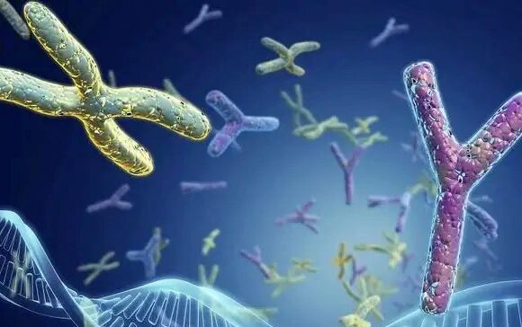
How do chromosomal abnormalities affect embryo quality?
I. Definition and classification of chromosomal abnormalities: the crisis of “genetic code” of embryo development
Chromosomal abnormality refers to abnormal changes in the number or structure of chromosomes, which is one of the core factors leading to embryonic development failure, birth defects and genetic diseases. Human cells contain 23 pairs of chromosomes (22 pairs of autosomes + 1 pair of sex chromosomes), and an imbalance in any one pair of chromosomes can lead to catastrophic consequences.

- Abnormalities in chromosome number
Aneuploidy:
Trisomy (e.g. trisomy of chromosome 21 → Down syndrome, trisomy 18 → Edwards syndrome)
Monosomal syndromes (e.g., X chromosome monosomy → Turner syndrome)
Polyploidy: e.g. triploidy (69 chromosomes), embryos usually do not survive to mid-gestation.
- Chromosomal structural abnormalities
Deletions (e.g. 5p deletion → cat-call syndrome)
Duplications (e.g. 16p11.2 duplication → increased risk of autism)
Translocations (carriers of balanced translocations may have chromosomally abnormal offspring)
Inversions (e.g., chromosome 9 inter-arm inversion may trigger recurrent miscarriages)
Ⅱ.The 5 major mechanisms by which chromosomal abnormalities affect embryo quality
- Early embryonic developmental stagnation: “fatal error” in gene expression.
About 50%-70% of early spontaneous abortions are directly related to chromosomal abnormalities in embryos. Abnormal chromosome combinations interfere with the following key processes:
Zygote genome activation (ZGA): on day 3 after fertilization, the embryo initiates its own gene expression, and the chromosomal abnormality results in the silencing of key genes (e.g., OCT4, NANOG).
Uncontrolled cell division: aneuploidy triggers failure of the spindle checkpoint (SAC), leading to multipolar division or division arrest.
Clinical data:
Only 20% of trisomy 21 embryos develop to the blastocyst stage;
Less than 5% of trisomy 16 embryos reach the blastocyst stage.
- Organogenesis disorders: “program collapse” of tissue differentiation.
Chromosomal abnormalities cause organ malformations by disrupting the HOX gene cluster (which regulates axial body development) and the WNT signaling pathway (which controls cell fate):
Heart defects: atrial septal defects are present in 40%-60% of children with trisomy 21;
Neurological abnormalities: failure of neural tube closure in up to 80% of trisomy 18 embryos.
- Impaired placental function: “nutritional cut-off” at the maternal-fetal interface.
Placenta-specific genes (e.g. CGA, PLAC1) are located in chromosomal regions prone to aneuploidy (e.g. 16, 22):
Triploid placenta: overgrown placental tissue triggers gestational hypertension and fetal growth restriction (FGR);
Uniparental diploidy (UPD): if chromosome 15 is UPD, it may induce Prader-Willi syndrome (placental hypofunction).
- Metabolic and epigenetic disorders: the “energy crisis” of cellular activity.
Mitochondrial dysfunction: trisomic cells require extra energy to maintain proliferation, leading to ATP depletion and oxidative stress;
Abnormal DNA methylation: genome-wide hypomethylation levels are 30% lower in Down syndrome embryos than in normal embryos.
- Immune rejection: maternally recognized “autoaggression”.
The maternal immune system recognizes the chromosomal status of the embryo through the HLA-G molecule:
Abnormal embryos have a 50% decrease in HLA-G expression, which triggers the activation of natural killer cells (NK cells);
Clinical statistics show that the maternal Th1/Th2 cytokine ratio is 3 times higher in chromosomally abnormal embryos, increasing the risk of miscarriage.
Ⅲ.Risk Factors for Chromosomal Abnormalities: A Comprehensive Analysis from Genetics to Environment
- Genetic factors
Balanced translocation carriers: the risk of having chromosomally abnormal offspring is 10-15%;
Roche translocation: the rate of abnormal embryos in people with chromosome 13/14 translocation is about 1%-2%.
- Maternal age effect
Egg aneuploidy rate:
20-25 years: 20%
35 years old: 40%
40 years: 80%
Mechanism: Loss of adhesin (Cohesin) with age during meiosis of the oocyte, leading to chromosome nondisjunction.
- Environment and Lifestyle
Radiation exposure: 1 Gy of radiation increases the risk of oocyte aneuploidy by a factor of 5;
Chemical pollutants: Bisphenol A (BPA) interferes with spindle assembly and triggers chromosomal errors in the egg;
Smoking: nicotine increases the rate of oxidative damage to oocytes 3-fold.
- Random mutations and gamete formation errors
Sperm aneuploidy rate: about 5%-10%, weak correlation with male age;
Chimerism: about 20% of embryos have intercellular differences in chromosome composition.
Ⅳ.Testing techniques for chromosomal abnormalities: a precise path from screening to diagnosis
- Non-invasive prenatal testing (NIPT)
Principle: analyze fetal free DNA (cfDNA) in maternal blood;
Detection rate: trisomy 21 >99%, trisomy 18 >97%, trisomy 13 >90%;
Limitation: unable to detect structural abnormalities and low percentage of chimeras.
- Pre-implantation Genetic Test (PGT-A)
Applicable people: repeated miscarriage, advanced age, balanced translocation carriers;
Technical advantage: Blastocyst stage biopsy + Whole Genome Sequencing (WGS), accuracy rate >98%;
Clinical value: increase the live birth rate of women over 35 years old by 20%-30%.
- Invasive diagnostic techniques
Chorionic villus sampling (CVS): performed at 10-13 weeks of pregnancy to detect chromosomal abnormalities;
Amniocentesis: performed at 16-20 weeks of pregnancy to diagnose open neural tube defects.
V. Response strategy: scientific reduction of the risk of chromosomal abnormalities
- Pre-conception intervention
Genetic counseling: karyotyping for those with family history or repeated miscarriages;
Antioxidant therapy: Coenzyme Q10 (600 mg/day) to improve egg mitochondrial function.
- Assisted Reproductive Technology (ART) optimization
Egg/embryo freezing: fertility preservation until 35 years of age;
Oocyte activation (ICSI-AOA): for high-risk couples with fertilization failure.
- Precision in embryo selection
Artificial Intelligence (AI) embryo assessment: prediction of chromosomally normal embryos by morphodynamics (85% accuracy);
Time-lapse imaging (TLI): dynamic monitoring of division rhythms to screen high-quality embryos.
- Future Technology Outlook
CRISPR gene editing: repairing chromosomal errors in embryos (still in the experimental stage);
Synthetic embryology: using stem cells to construct artificial embryos to circumvent genetic defects.
VI. Conclusion: the leap from mechanism knowledge to clinical practice
The mechanisms by which chromosomal abnormalities affect embryo quality are complex and multidimensional, but modern reproductive medicine has provided a complete solution from prevention, detection to intervention. Through the precise application of technologies such as PGT-A, combined with lifestyle optimization and genetic counseling, the pregnancy risk of abnormal embryos can be significantly reduced. In the future, with the development of gene editing and artificial intelligence, human beings will be more proactive in controlling the “genetic code” of embryo health.
Search within the site
Surrogacy News
Hot Tags.
Kyrgyzstan Surrogacy Agency,Global IVF Hospitals,International Surrogate Mother Recruitment