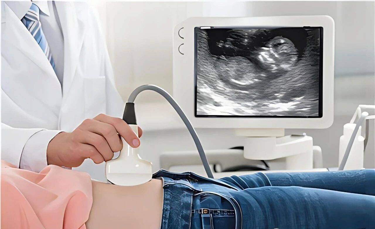
IVF Early Pregnancy Screening
From the fertility clinics on the Left Bank of Paris to the Fetal Medicine Center in Manhattan, New York, a quiet revolution is taking place in the global field of assisted reproduction – moving the screening gate for major malformations forward to 11-13 weeks of pregnancy. For IVF families, this revolution is not only a breakthrough in medical technology, but also a strategic shift in guarding the quality of life.
Chapter 1: NT Screening – The First Safety Alert of Life
The Truth Revealed by Data
An analysis of 560,000 pregnancy cases by the Danish National Institute of Health shows:
21% of fetuses with NT ≥ 3.5mm had chromosomal abnormalities
At NT ≥ 6.5mm, the rate of chromosomal abnormality soared to 69%
Even if chromosomally normal, the rate of healthy live births in fetuses with thickened NT decreases sharply with increasing thickness:
3.5-4.4 mm: 87%
≥6.5 mm: 29%
Scientific Metaphor
The NT test acts as a “biological radar” for the embryo, where a difference in thickness of 2.5 mm can mean the difference between a healthy life and a major defect.
International case studies
Sophie (32) from Berlin, Germany, had an NT of 4.2 mm at 12 weeks after an IVF cycle, and despite normal chromosomal testing, fetal medicine specialist Dr. Müller recommended ongoing monitoring, which eventually led to the discovery of a complex cardiac abnormality at 18 weeks of pregnancy. “NT thickening is the first warning letter sent by life, and it takes wisdom and courage to interpret it.” Sophie exclaimed at the post-operative sharing session.
Chapter 2: The Chromosome Puzzle – Deep Screening Beyond Traditional Perceptions
Technological revolution: from karyotyping to chromosome microarray (CMA)
Detection accuracy: CMA can identify microdeletions/duplications of 5-10Mb, 100 times more sensitive than traditional karyotype analysis
Limitation breakthrough:
Cambridge, UK team finds that 12% of CMA normal fetuses still have epigenetic abnormalities
University of Tokyo, Japan, Develops AI Prediction Model Combining NT and Metabolomics Data to Increase Risk Prediction Accuracy to 89 Percent
Chapter 3: Cardiac Screening – A Technological Miracle with a 0.01% Misdiagnosis Rate
The Weight of Life Behind the Numbers
Global status: congenital heart disease accounts for 28% of birth defects, with 70-80 cases per 10,000 births
Screening Breakthrough:
Ultrasound sensitivity of 70.5% and specificity of 99.99% in early IVF pregnancy (70,000 population study)
Four-dimensional flow imaging captures ventricular septal defects at 0.2mm level
Interdisciplinary collaborative model
Embryologists: Evaluating the cardiac developmental potential of IVF embryos
Fetal Cardiologist: Dynamic Monitoring Initiated at 11 Weeks of Pregnancy
Genetic Counselor: Interpretation of WES (Whole Exome Sequencing) Reports
Innovative Tool:
FDA Approves AI Heart Modeling System to Predict Malformation Risk from NT Data
Germany Develops Miniature Ultrasound Probe to Transvaginally Detect Fetal Heart Tube Beats at 9 Weeks of Pregnancy
Chapter 4: The Time Game – The Strategic Value of 10-Week Pregnancy Screening
The Clinical Significance of Moving the Alarm Forward
Key finding: those with NT ≥2.5mm at 10 weeks’ gestation had an 8.5% risk of adverse outcomes even with normal values at 11 weeks’ gestation
Response strategy:
Establish an “early warning system”: initiate nuchal translucency monitoring at 9 weeks of gestation.
Developing dynamic NT tracking algorithm to predict extrachromosomal abnormalities.
Upgrading of international standards of care
| gestation period | Inspection items | technical target |
|---|---|---|
| 9-10 weeks | Transvaginal NT + Flow Doppler | Recognizing Early Signs of Heart Failure |
| 11-13 weeks | Standard NT + Fetal Nasal Bone Assessmentv | Primary screening for chromosomal abnormalities |
| 14-16 weeks | Non-invasive DNA + echocardiogram | Depth screening for structural deformities |
Chapter 5: An Action Guide for IVF Families Worldwide
The Three-Tier Defense System
Embryo Selection:
PGT-A Screening to Exclude Chromosomal Aneuploidy
Time-lapse imaging system to assess embryo morphodynamics
Early Ultrasound Monitoring:
Transvaginal ultrasound at 9 weeks of gestation (requires specialized fetal medicine center)
Combined serum PAPP-A + β-hCG screening
Dynamic tracking:
Establishment of NT change curve (weekly testing from 9-13 weeks of gestation)
Complete whole exome sequencing by 18 weeks of gestation
Environmental optimization program
Nutritional intervention:
Folic acid (800μg/day) + vitamin B12 (4μg/day) to reduce the risk of neural tube defects
Omega-3 fatty acids (DHA 200mg/day) to promote fetal heart development
Toxin Defense:
Use glass containers instead of plastic
Installation of reverse osmosis water purification systems (removes nanoplastics)
Chapter 6: Future Prospects – Redefining the Beginning of Life
Dawn of Gene Editing
CRISPR Technology: U.S. Team Successfully Repairs Cardiac Malformation Gene in Mouse Model
Mitochondrial Replacement: UK Approves “Three-Parent IVF” Technology to Block Maternal Genetic Diseases
Life Lessons
IVF early pregnancy screening is not a simple medical process, but a deep conversation between human beings and the mysteries of life. From one-millimeter fluctuations in NT values to precision imaging of heart structures, technology is transforming the uncharted territory of fertility into a quantifiable scientific path. As Nobel Prize winner Jennifer Doudna said, “We are learning to negotiate life in the language of molecules.” On this path, every careful screening is the most solemn commitment to life.